Research Overview
Structure and the biocytin linkage
Biotin's two fused rings supply the ureido nitrogen that carries an activated carboxyl group during catalysis, while the valeric acid tail tethers the vitamin to its enzyme. Holocarboxylase synthetase forms an amide bond between that tail and a specific lysine on each apocarboxylase; biotinidase later cleaves biocytin during protein turnover, recycling the vitamin. Defects in either enzyme are the basis of the multiple carboxylase deficiency models studied in metabolic biochemistry.
Carboxylase biochemistry
- Acetyl-CoA carboxylase 1 and 2 — malonyl-CoA production and the regulation of fatty acid oxidation
- Pyruvate carboxylase — the anaplerotic entry to gluconeogenesis
- Propionyl-CoA carboxylase — odd-chain fatty acid and branched amino acid catabolism
- Methylcrotonyl-CoA carboxylase — leucine degradation
Biotin-streptavidin as a research tool
Assay interference
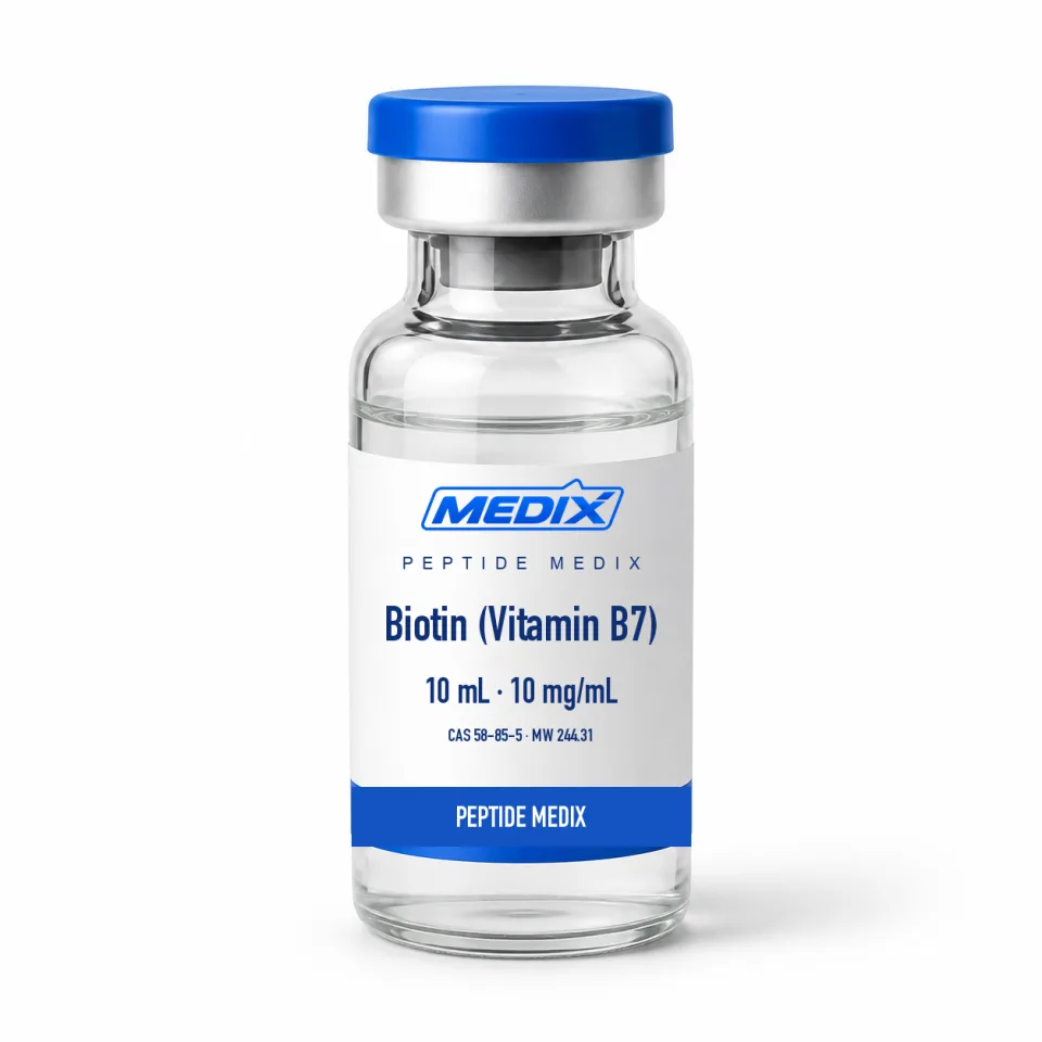
Concentration verification is also worth building into the workflow. Biotin has no strong chromophore in the visible range and only weak ultraviolet absorbance, so spectrophotometric quantification is unreliable; HPLC or a competitive binding assay against a labelled tracer gives a far more dependable measure of the free vitamin present.