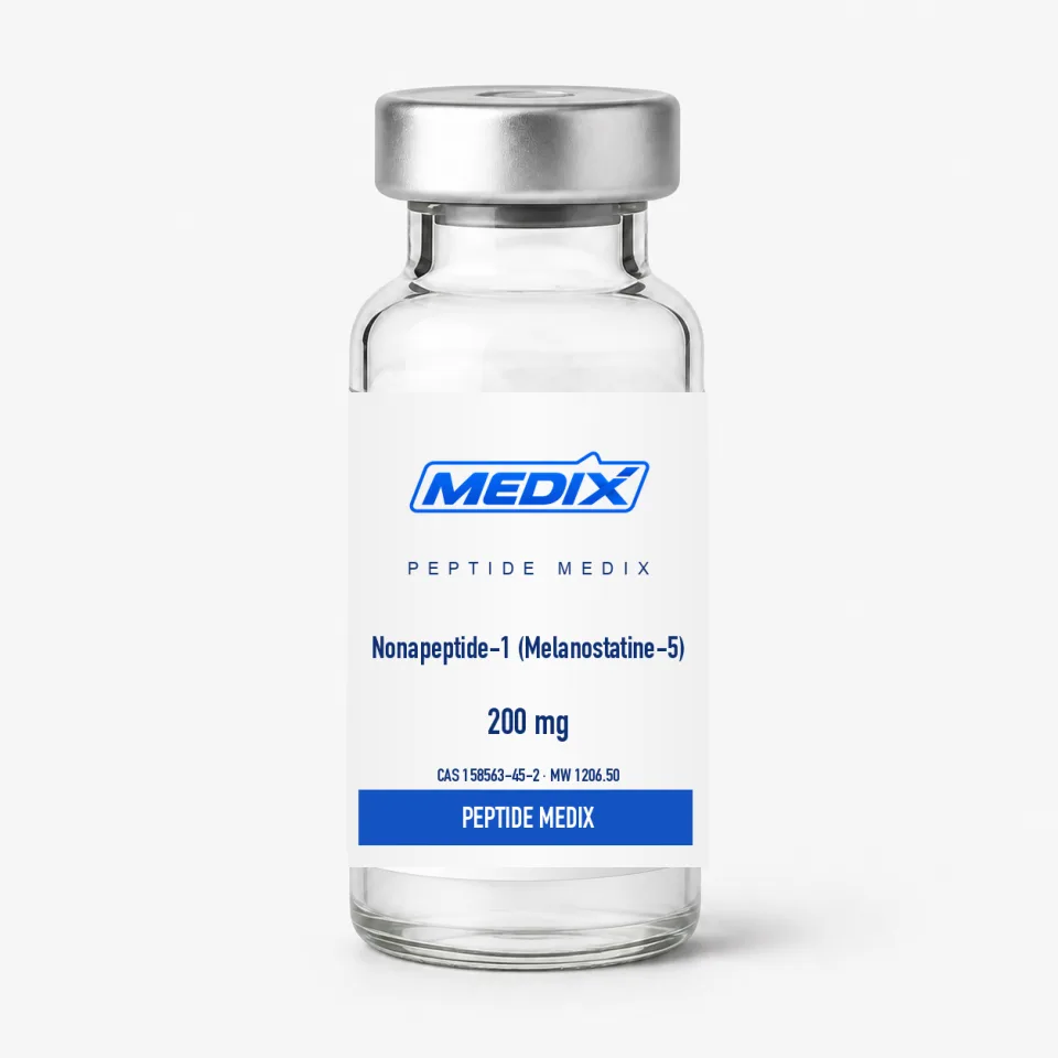
Research Overview
MC1R pathway placement
Alpha-melanocyte-stimulating hormone activates MC1R on melanocyte models, raising cyclic AMP and increasing transcriptional programs that include tyrosinase. The canonical route runs through protein kinase A and CREB to the microphthalmia-associated transcription factor, which in turn governs tyrosinase and the tyrosinase-related proteins. Nonapeptide-1 was selected as an antagonist candidate against this receptor axis. It is therefore used upstream of pigment synthesis, unlike a direct tyrosinase inhibitor that acts on the enzyme itself, and antagonism is normally reported as suppression or a rightward shift of an agonist concentration-response curve rather than as an effect measured on its own.
Sequence design
The D-Phe and D-Trp residues are not typographic details. Reversing stereochemistry at those positions changes backbone geometry and resistance to common proteases, most of which are stereospecific for L residues. The C-terminal amide also changes charge relative to a free carboxylate. Identity testing should be able to distinguish the intended stereochemical and terminal form rather than reporting only nominal mass: the amide and the free acid differ by roughly one dalton, and stereochemistry is invisible to mass spectrometry altogether, so chiral amino-acid analysis or a resolving chromatographic method is what settles the question.
Typical experimental readouts
- Ligand competition or functional antagonism in MC1R-expressing cells, often against a labelled superpotent agonist such as NDP-alpha-MSH
- Intracellular cyclic-AMP response after alpha-MSH stimulation
- Tyrosinase activity and expression in melanocyte or melanoma-cell models, commonly read as L-DOPA oxidation
- Melanin content in cultured cells and reconstructed pigmented tissue
- Selectivity counter-screens at the other melanocortin receptors
- Stability comparisons between the D-amino-acid peptide and all-L controls
Controls a study should carry
Forskolin or a phosphodiesterase inhibitor raises cyclic AMP downstream of the receptor, so a compound that blunts the alpha-MSH response while leaving the forskolin response intact is acting at or near MC1R rather than further down the cascade. Reference inhibitors such as kojic acid or arbutin anchor the pigment readout, and vehicle-matched, density-matched wells are what make small pigment differences interpretable.
Interpretation limits
Melanogenesis models are sensitive to cell source, baseline pigment, passage number, UV exposure and vehicle. Reduced pigment readouts can also result from cytotoxicity, so viability and morphology controls are essential. Murine melanoma lines and human melanocytes do not share melanocortin pharmacology exactly, and MC1R is polymorphic in human populations, with common variants reported to signal less efficiently. Nonapeptide-1 should be compared with Decapeptide-12 only with the understanding that the two molecules act at different points in the pathway.