Research Overview
Discovery and family
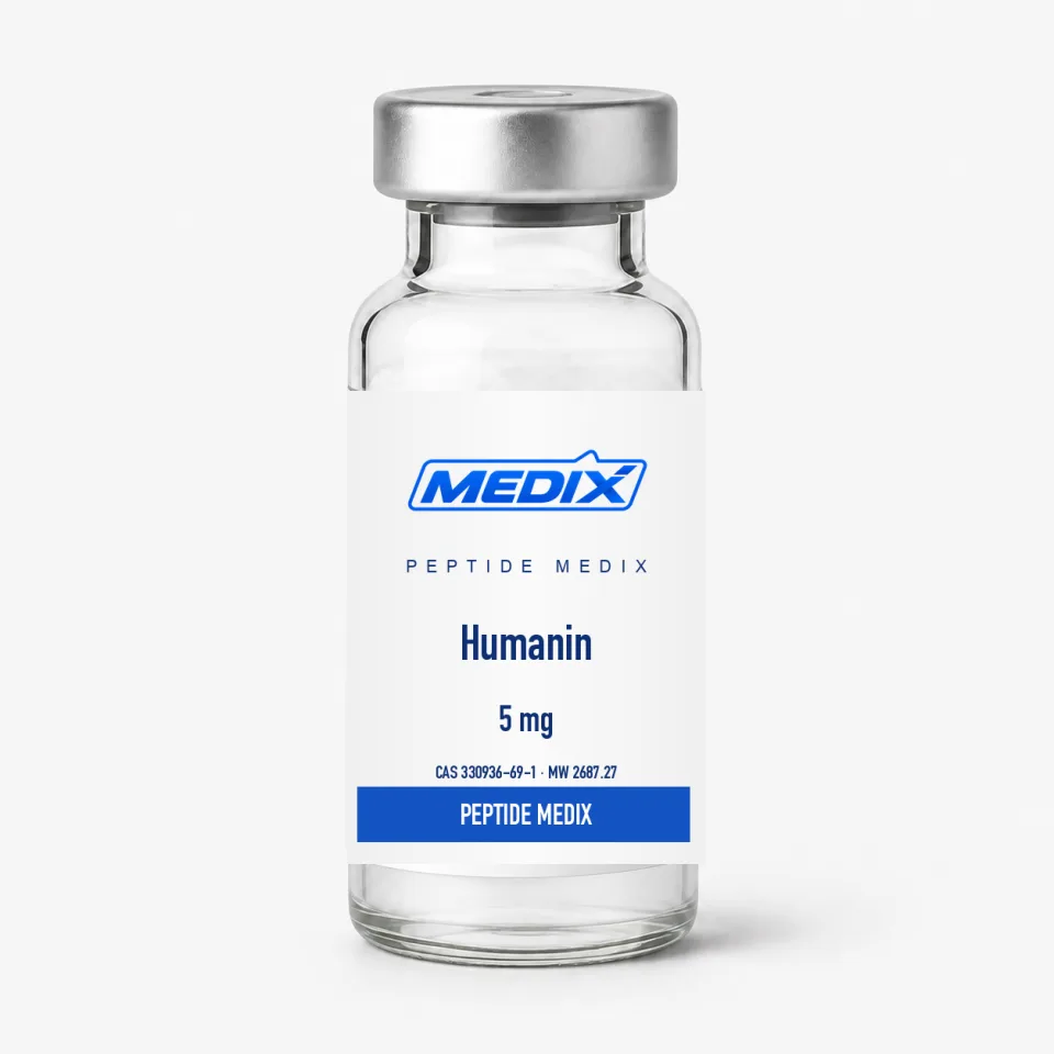
Humanin emerged from a functional screen rather than a sequence prediction, and its identification opened the field of mitochondrial-derived peptides that later grew to include MOTS-c and the SHLP series. The open reading frame sits inside the 16S rRNA gene; both mitochondrial and nuclear-encoded variants have been reported, and several analogues such as HNG (S14G-humanin) appear widely in the literature as more potent research tools.
Reported signaling interactions
- Binding to the pro-apoptotic protein Bax and interference with its mitochondrial translocation in cell models
- Association with IGFBP-3, an interaction described in early biochemical studies
- Engagement of a trimeric receptor complex (CNTFR / WSX-1 / gp130) linked to STAT3 activation
- Reduced markers of apoptosis in serum-deprived and stress-challenged cultures
Animal studies have examined humanin in models of neurodegeneration, ischemia, atherosclerosis and insulin resistance, generally reading out cell survival, inflammatory markers and glucose handling. Observational work reports that circulating levels decline with age in humans and rodents and are elevated in some long-lived mouse models, which is why the peptide appears frequently in aging biomarker studies. These findings describe associations in research settings and are not statements about outcomes in people.
Chemistry and handling considerations
The central Leu-Leu-Leu-Leu stretch makes humanin relatively hydrophobic for its length, and preparations occasionally need gentle warming or a small volume of a compatible co-solvent to dissolve fully. The methionine at position one and the single cysteine are the residues most sensitive to oxidation, so light protection and cold storage matter for long experiments.