Research Overview
Why PEGylation is used
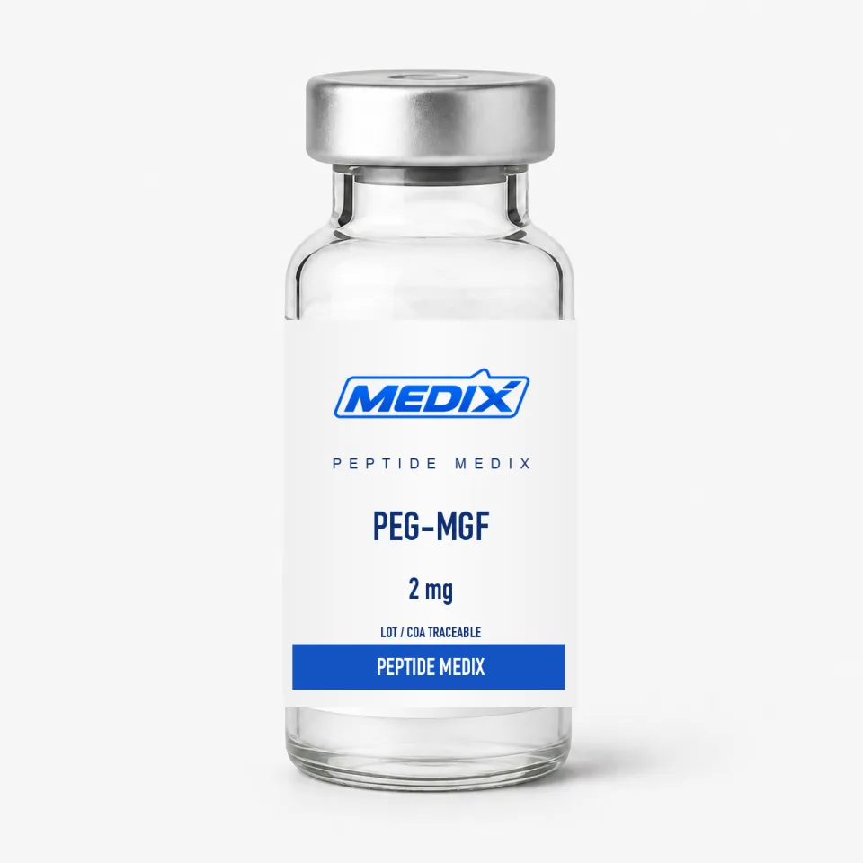
Small peptides are cleared quickly: they are filtered by the kidney and cut by circulating proteases within minutes. Covalently attaching polyethylene glycol adds bulk and a shell of associated water that physically obstructs enzymatic access and pushes the molecule above the renal filtration threshold. The approach is well established across protein pharmacology, and PEG-MGF is a straightforward application of it to the MGF E-domain.
- Direct comparisons of PEGylated and unmodified MGF exposure profiles in rodent serum and tissue.
- Myoblast and satellite-cell proliferation assays where the extended exposure window changes the observed response curve.
- Models of muscle crush injury, disuse atrophy and post-load remodelling, where investigators ask whether sustained E-domain presence alters the repair timeline.
- Cardiac tissue models, following reports that the same splice variant is induced after ischaemic stress.